Endoscopic Ultrasound, EUS, Endoscopic Ultrasound Purpose, Endoscopic Ultrasound Procedure, Endoscopic Ultrasound Results, Endoscopic Ultrasound Risks, Endoscopic Ultrasound Benefits
Endoscopic Ultrasound (EUS)- Purpose, Procedure and Results
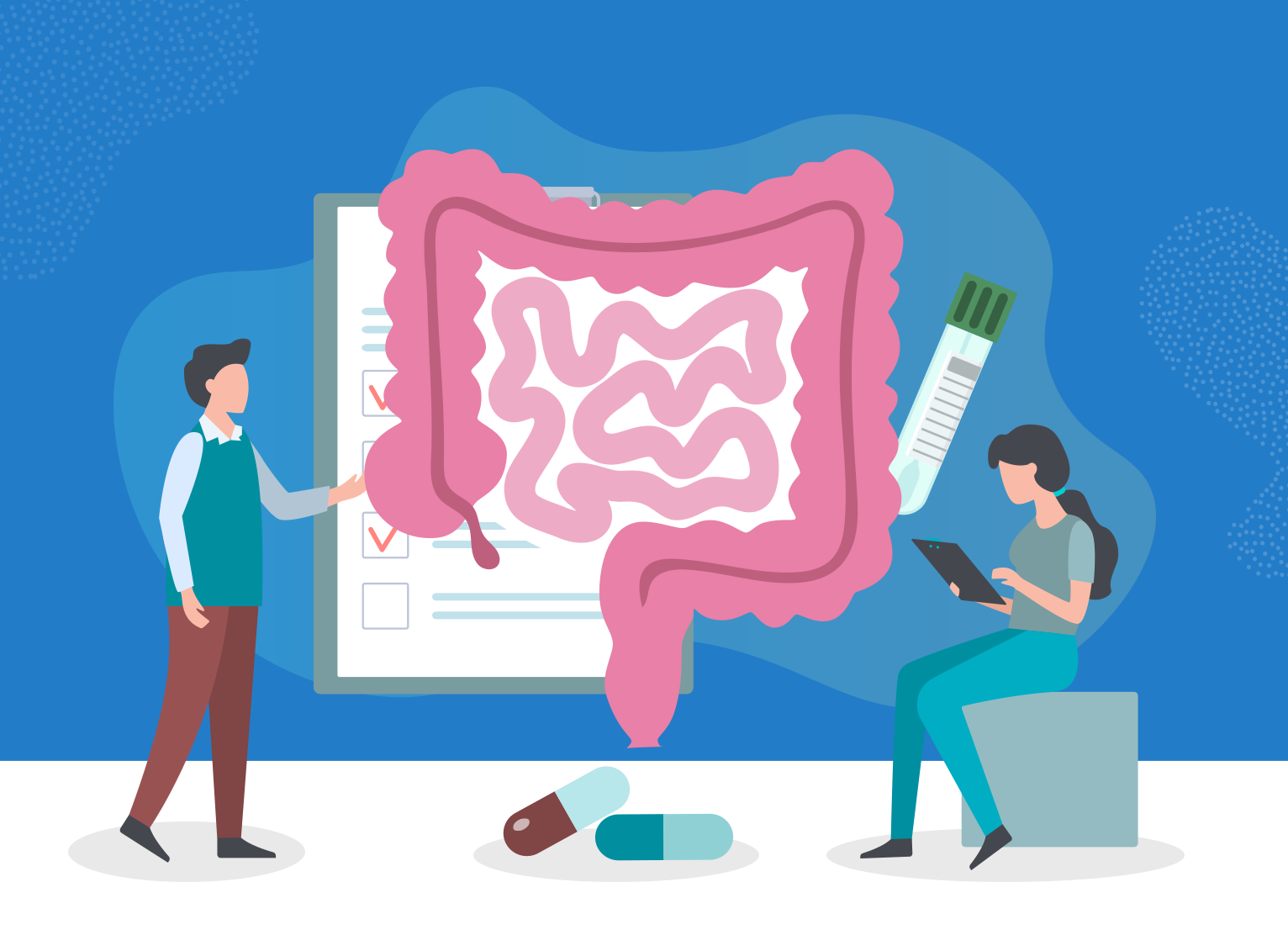
Endoscopic ultrasound (EUS) combines two techniques- endoscopy and ultrasound- to help doctors see, evaluate and diagnose conditions in and near the gastrointestinal (GI) tract.
What is an Endoscopic Ultrasound?
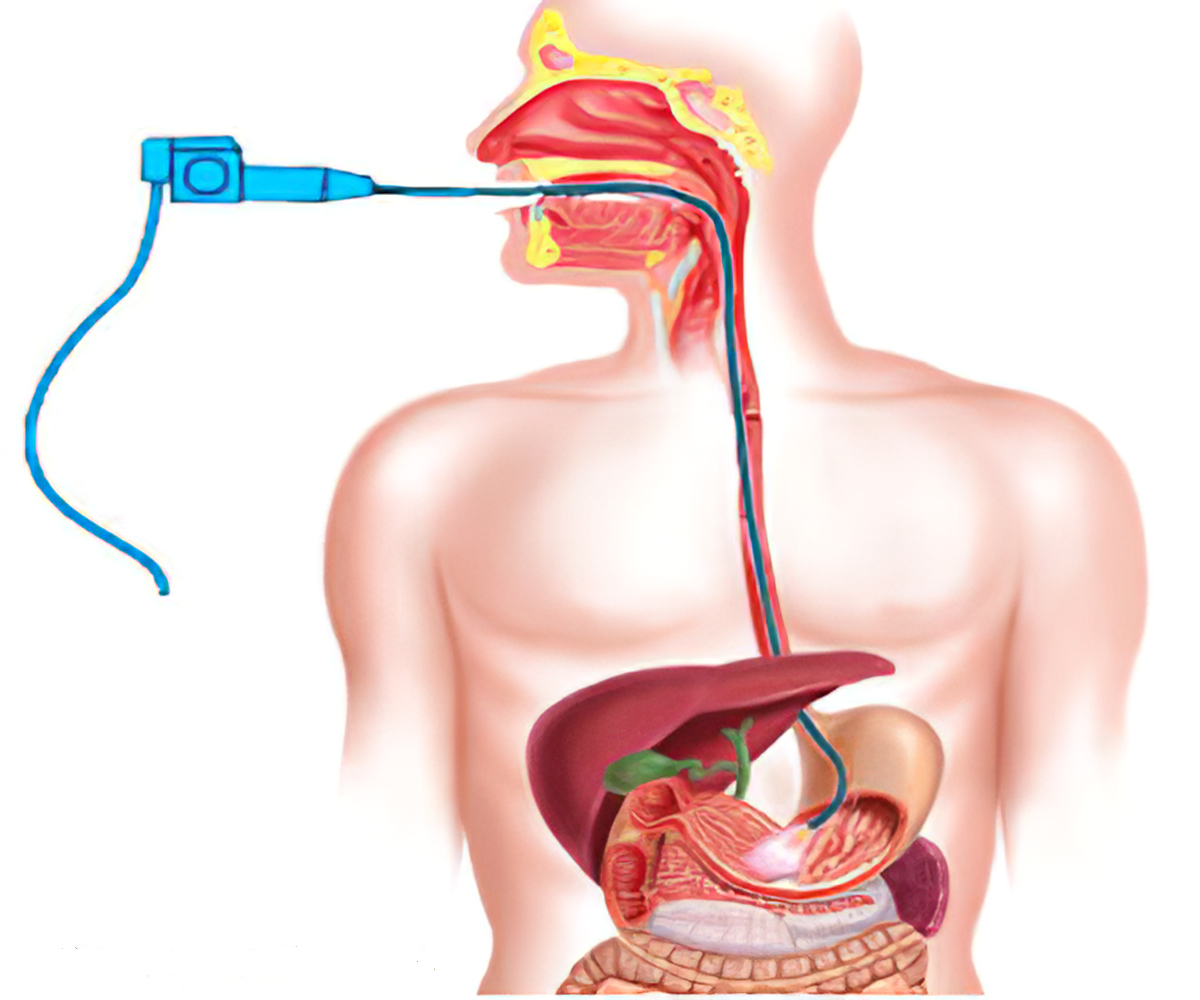
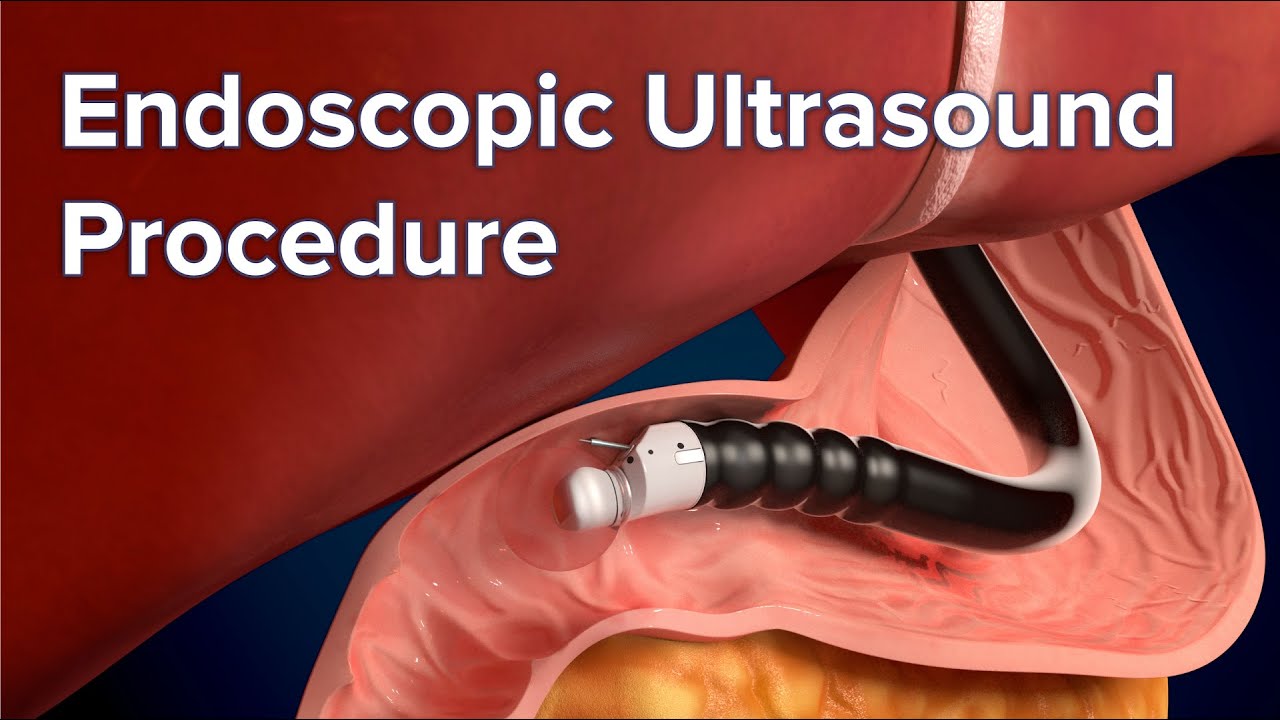
Endoscopic ultrasound is a minimally invasive procedure that uses a camera device (endoscope) together with high frequency sound waves (ultrasound) to examine the GI tract and beyond.
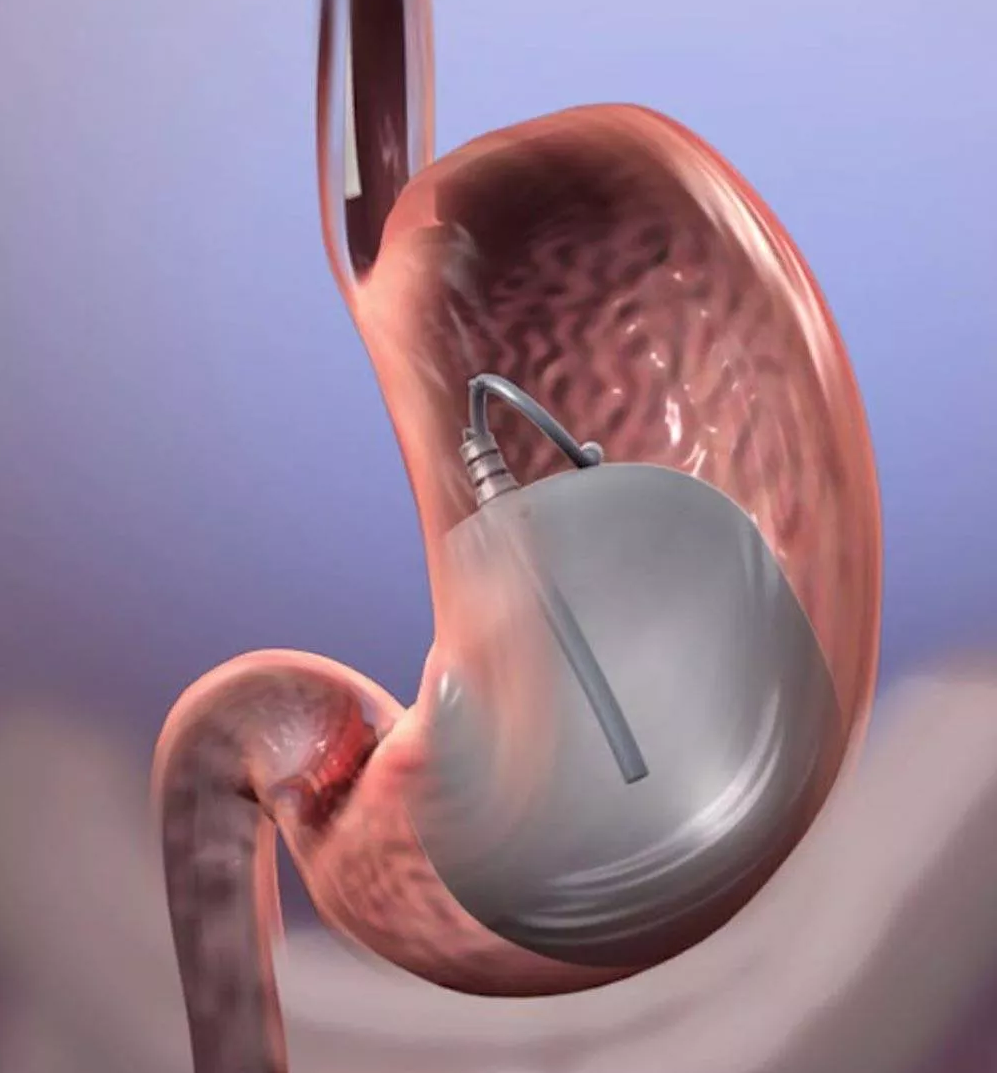
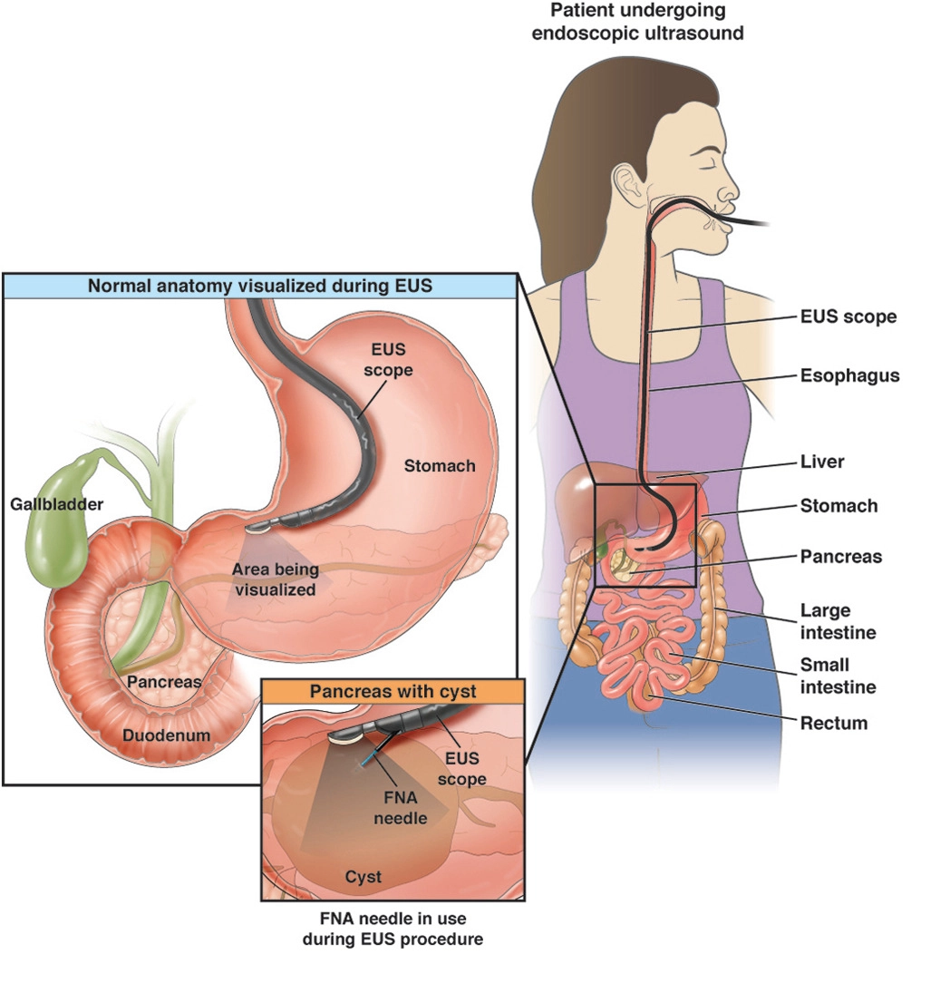
A regular endoscope is a thin, lighted tube that can be inserted through the mouth or anus to view inside your esophagus, stomach or intestines. EUS uses a special endoscope with a small ultrasound device on its tip, called an echoendoscope. Addition of ultrasound lets the doctor see the GI tract and surrounding organs.
By inserting the endoscope and camera into the upper or the lower digestive tract, the doctor is able to obtain high-quality ultrasound images of organs. Because the EUS can get close to the organ(s) being examined, the images obtained with EUS are often more accurate and detailed than images provided by traditional ultrasound which must travel from the outside of the body.
Why is Endoscopic Ultrasound done?
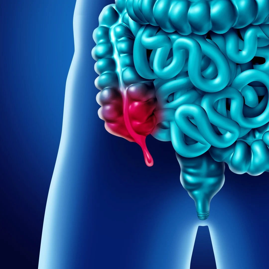
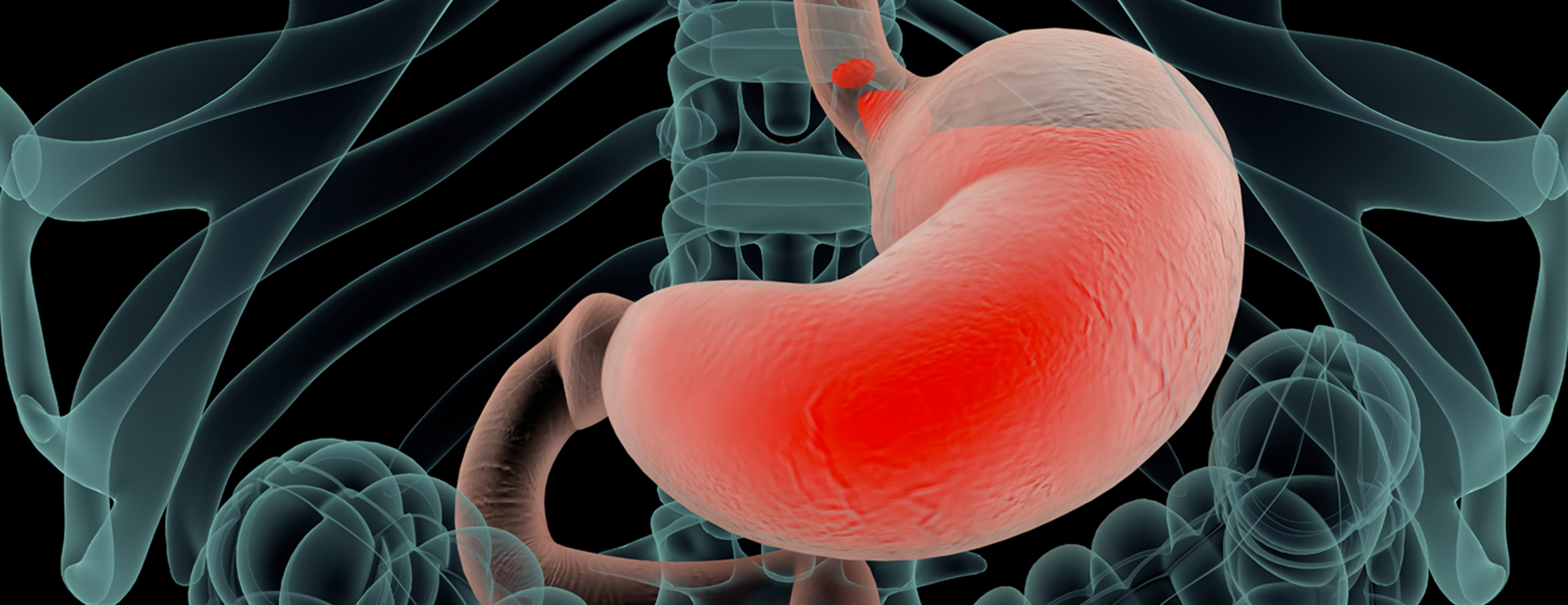
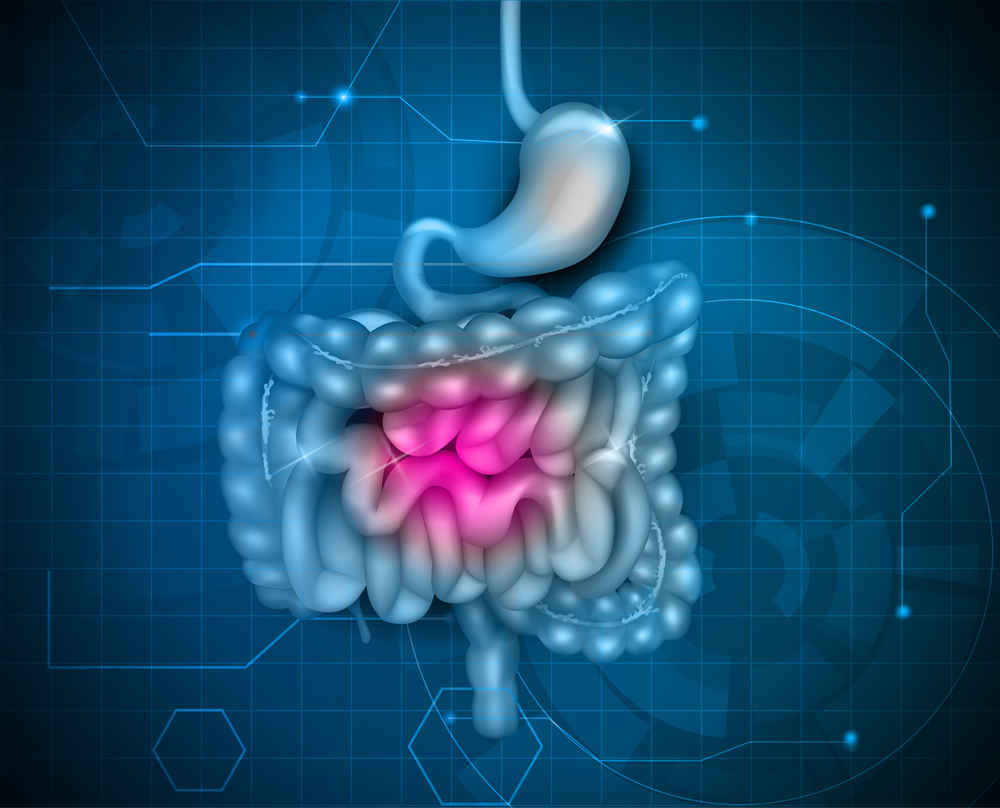
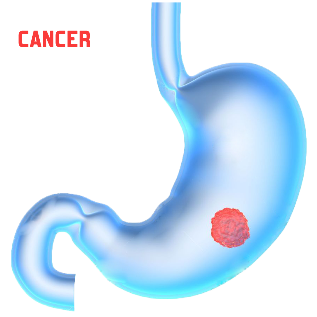
EUS can create images of the lining and walls of any organ along the route of the gastrointestinal tract. An EUS device placed down the throat can capture images of the esophagus, stomach and parts of the small intestine. An EUS device inserted through the anus captures images of the rectum, parts of the large intestine (colon) and surrounding tissue such as lymph nodes.
Internal organs or nearby structures can also be visualized, including the following:
- Lungs
- Lymph nodes in the center of the chest
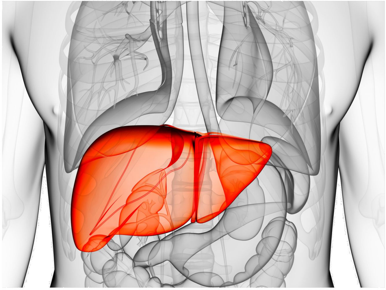
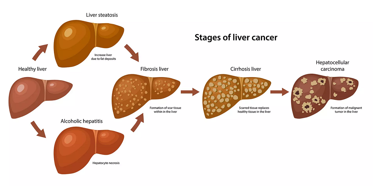
- Liver
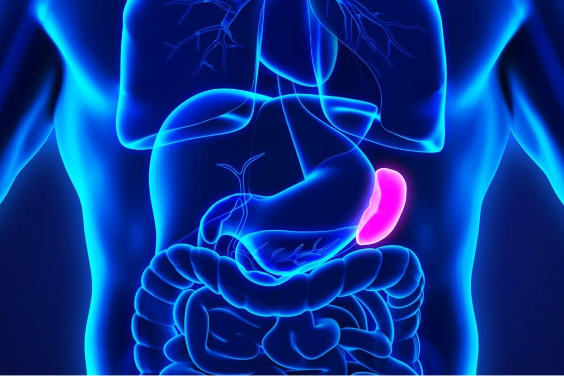
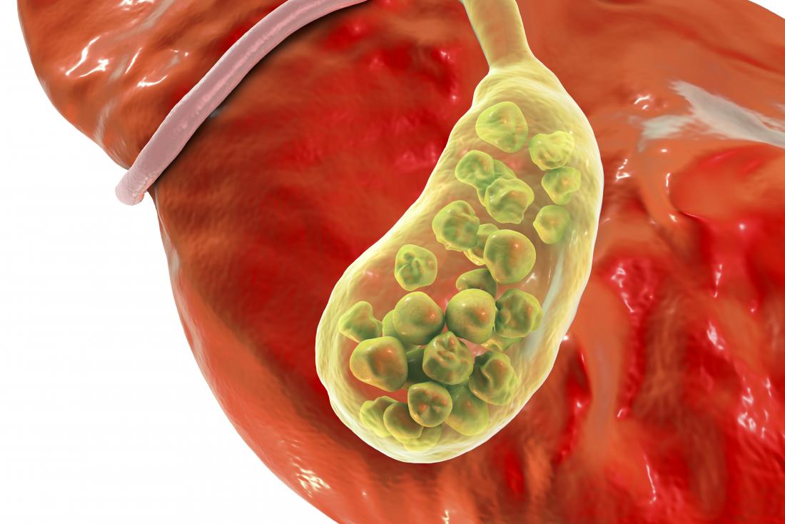
- Gallbladder
- Bile ducts
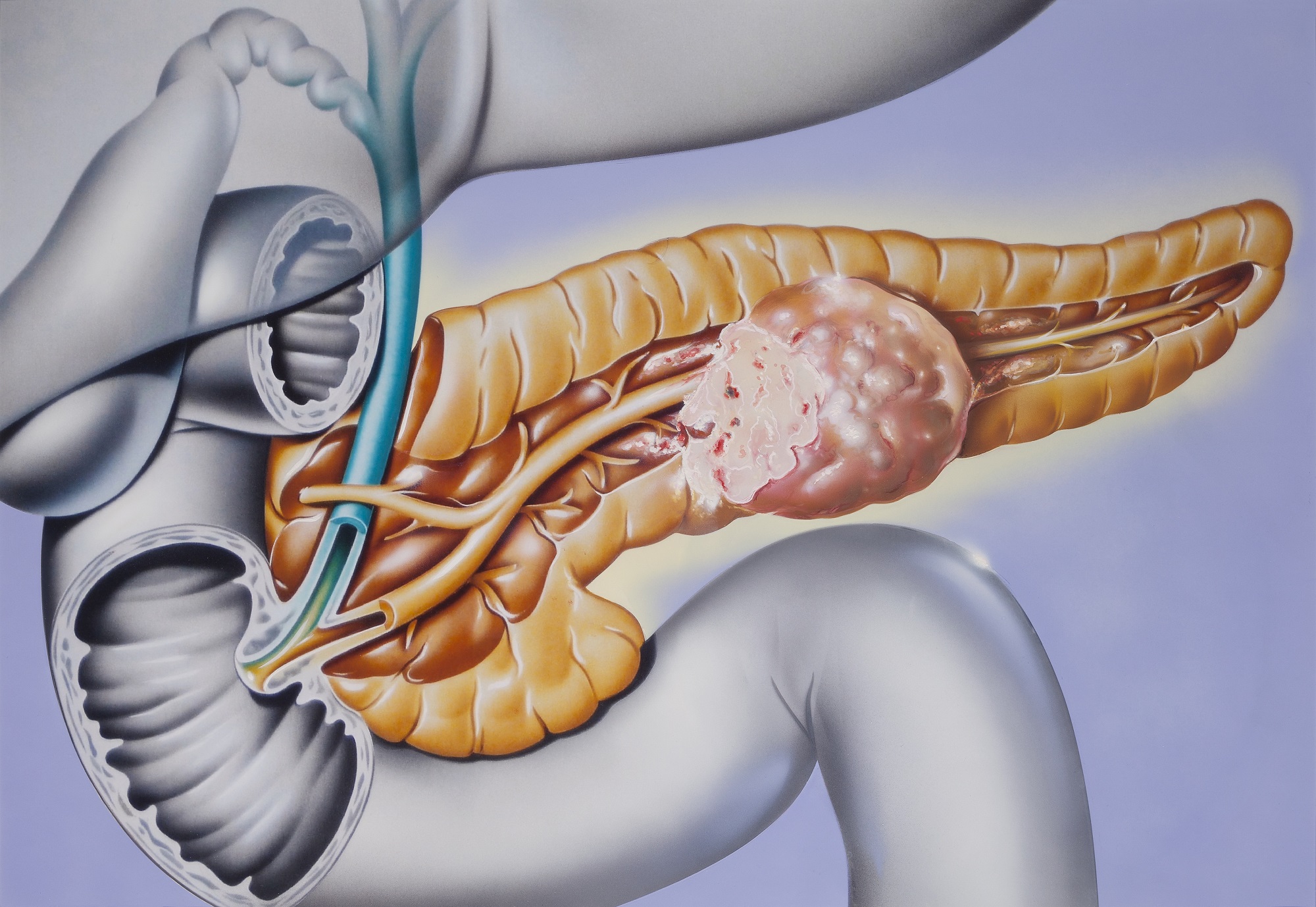
- Pancreas
EUS with fine-needle aspiration or other instruments can also reach these other organs. For example, a needle can pass through the wall of the esophagus to nearby lymph nodes. Similarly, a needle can pass through the wall of the stomach to deliver medication to the pancreas.
EUS and EUS-guided procedures can be used for the following:
- Assess damage to tissues due to inflammation or disease
- Determine whether cancer is present or has spread to lymph nodes
- Assess how much a cancerous (malignant) tumor invades tissues
- Determine how advanced cancer is
- Provide more-detailed information about lesions identified with other imaging technologies
- Extract fluid or tissue for diagnostic testing
- Drain fluids from cysts
- Deliver therapies to a targeted region, such as a malignant tumor
How does Endoscopic Ultrasound work?
Endoscopic ultrasound works similar to traditional ultrasound, except the source of the sound waves is inside your body. As the high frequency sound waves travel from the echoendoscope, they hit tissues of various densities and bounce back. For example, endoscopic ultrasound can pick up outlines of a tumor or a cyst, which the computer then processes and displays as lighter and darker areas on the screen.
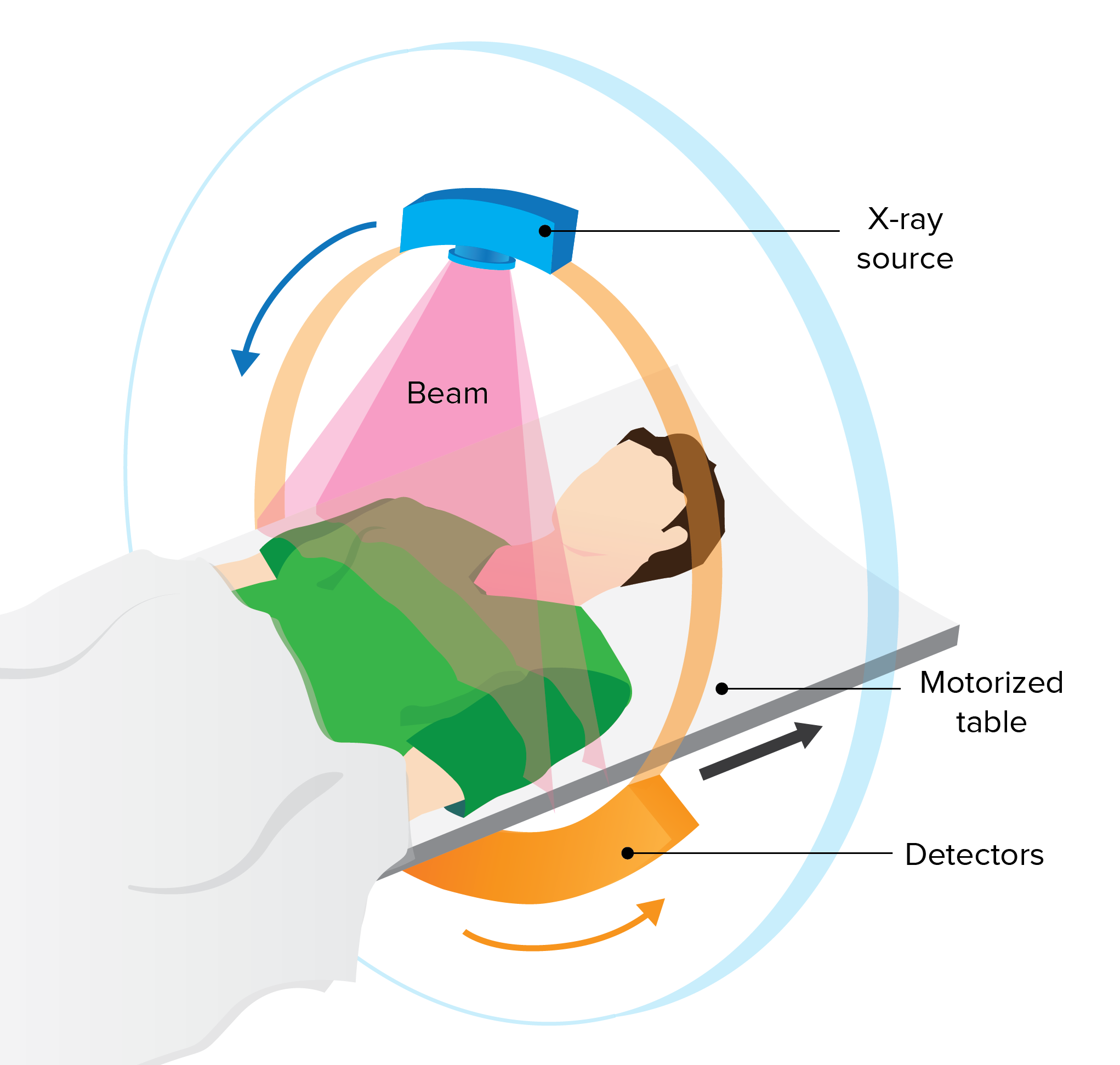
Because the sound waves don’t need to pass through the skin and muscle to reach internal organs, endoscopic ultrasound offers a better view of the GI tract and nearby organs than regular abdominal ultrasound. It may also be more precise in identifying small tumors and cysts that other imaging methods such as MRI (magnetic resonance imaging) and CT (computerized tomography) can miss.
During EUS, the gastroenterologist can take a tissue sample (biopsy) of a suspicious lesion by using a thin needle fed through the echoendoscope. This procedure, called fine-needle aspiration, can be used to collect cells and tissues from the pancreas, liver and other organs adjacent to the GI wall. Biopsy can determine the presence of cancer and examine its characteristics.
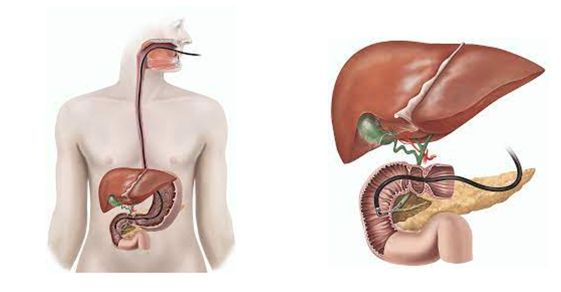
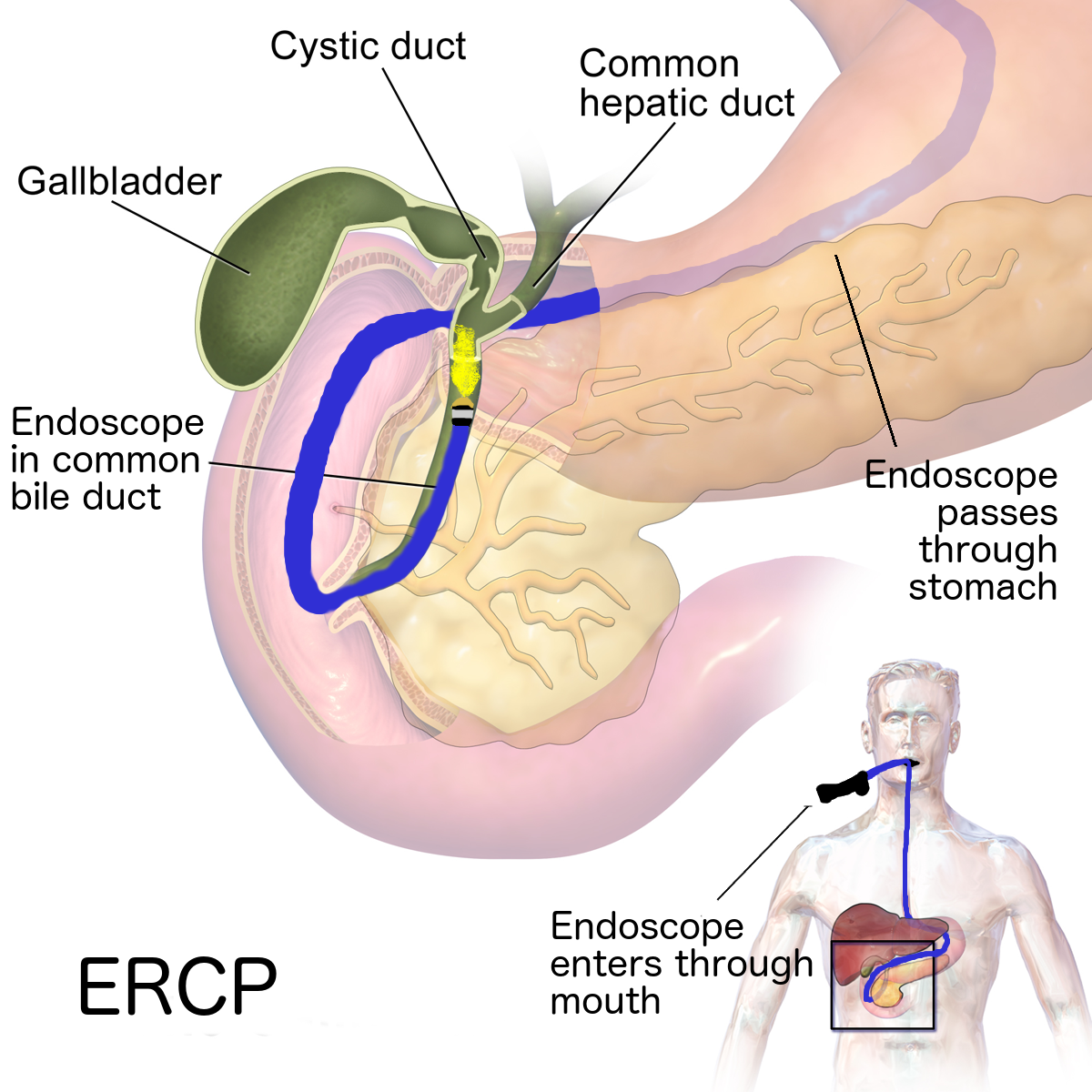
What are the advantages of an EUS compared to a CT Scan, MRI, Or ERCP?
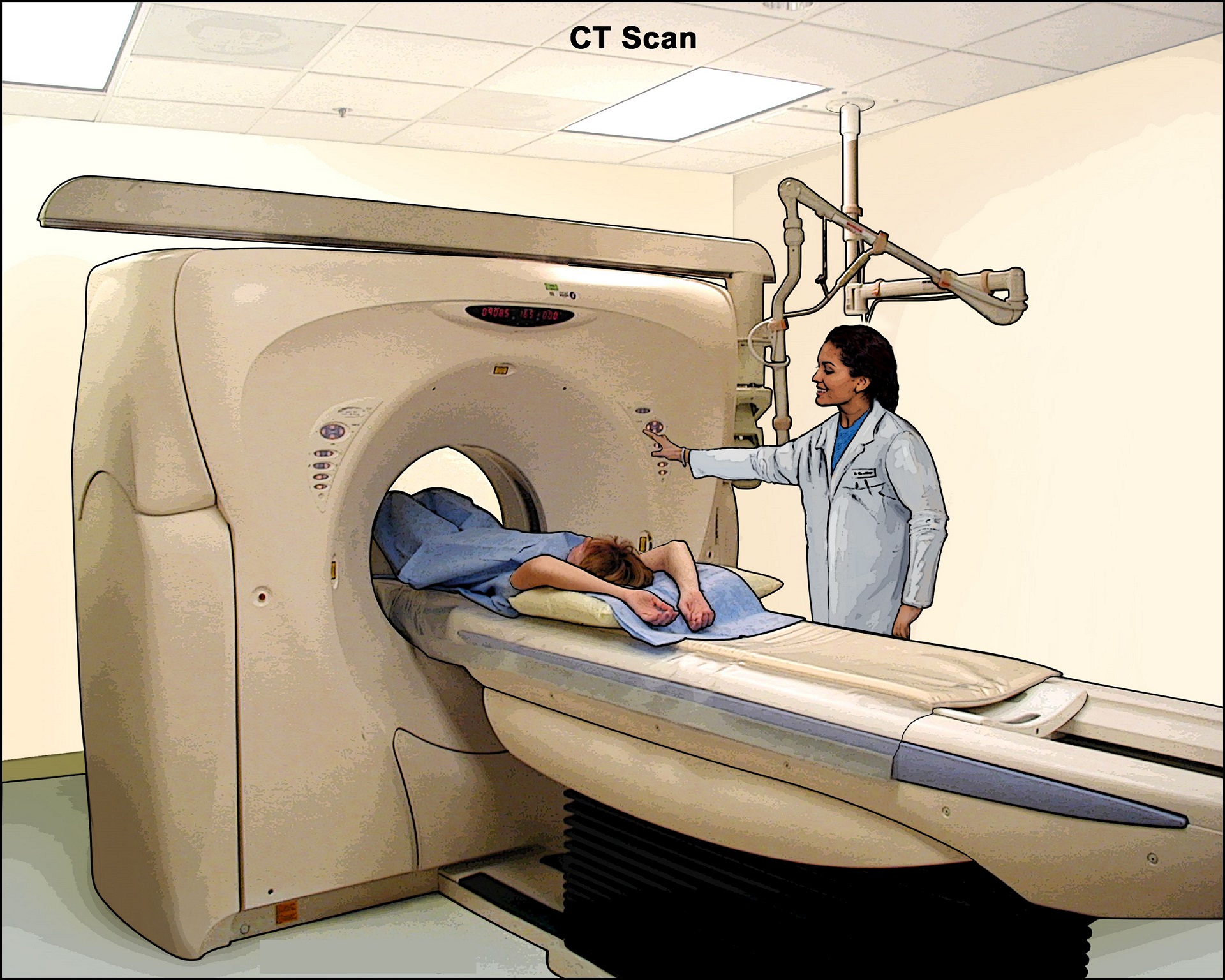
There are many different tests which can be used to evaluate the GI tract. CT scans and MRI are types of non-invasive tests which allow for detailed imaging of the organs and the surrounding structures in the abdominal cavity. CT scans expose the patient to some amount of radiation. Furthermore, some patients are unable to receive IV contrast for their CT scans (due to allergies or kidney problems), and thus the quality of the pictures will be sub-optimal. A special kind of MRI called an MRCP can give high-quality pictures of the organs, however, some patients who are claustrophobic may decide against having an MRI performed.
As discussed, EUS allows the physician to get in very close proximity which results in very detailed imaging of the organ. The endoscopist can often times visualize details of the organs that cannot be seen with either CT or MRCP. Furthermore, there is no exposure to radiation and no need for contrast to be given.
The biggest advantage of EUS is that, unlike with CT or MRCP, pancreatic biopsies can be safely and easily obtained at the time of the exam.
What are the risks of Endoscopic Ultrasound?
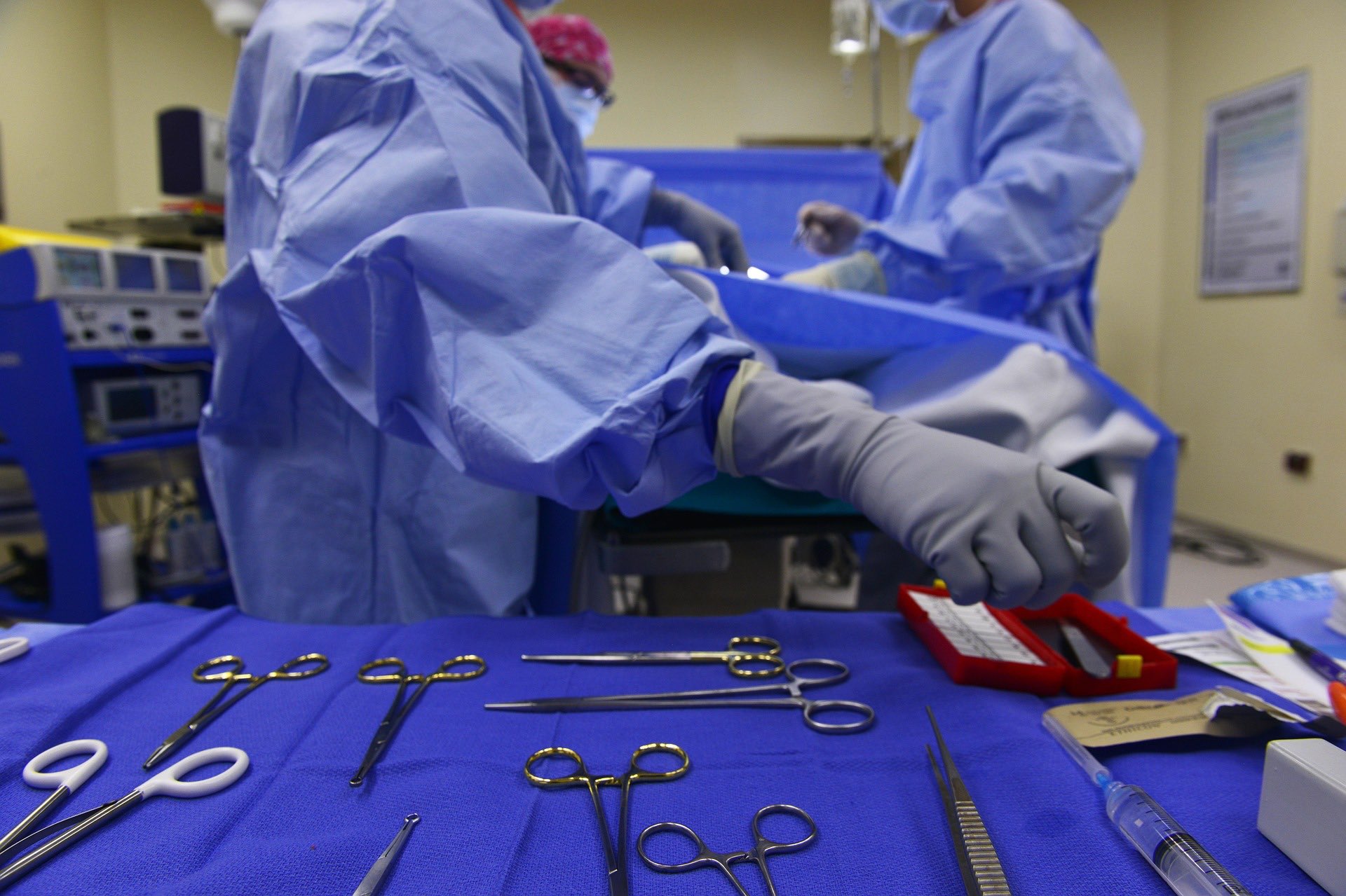
EUS is generally safe when performed at a centre with an experienced and expert health care team. The procedure is usually performed by a medical doctor who specializes in the digestive system (gastroenterologist) and has specific training in EUS procedures.
Your doctor will discuss with you the risk of complications from EUS. These risks, which are most commonly associated with fine-needle aspiration, may include:
- Bleeding
- Infection
- Tearing (perforation) of an organ wall
- Pancreatitis, if fine-needle aspiration of the pancreas is done
You can reduce your risk of complications by carefully following your doctor's instructions for preparing for EUS.
Call your healthcare provider immediately or go to an emergency room if you experience any of the following signs or symptoms after the procedure:
- Fever
- Severe or persistent abdominal pain
- Neck or chest pain
- Severe nausea or vomiting
- Vomiting blood
- Black or very dark-colored stool
How to prepare for Endoscopic Ultrasound
Your doctor will give you specific instructions to prepare for your EUS. Instructions will include the following:
- Fasting. You'll likely be asked to fast for at least six hours before the test to ensure your stomach is empty.
- Colon cleansing. If the EUS will be performed through the rectum, you'll be directed to use a colon cleansing solution or to follow a liquid diet and use a laxative before the procedure.
- Medication. Tell your doctor about all prescription and nonprescription medications you take, as well as herbal remedies and dietary supplements. Follow the instructions on which medications you can take and which you need to stop taking before the procedure.
- Travel. Medication to help you relax or sleep (sedative) or anesthesia can impair your coordination and judgment after the procedure. Arrange for someone to drive you home and stay with you the rest of the day.
What to expect during Endoscopic Ultrasound
If you are given anesthesia, you won't be conscious during the procedure. If you're given a sedative, you may feel a slight discomfort, but many people fall asleep or are not alert during the procedure.
You'll likely lie on your left side during the procedure. The doctor feeds a thin, flexible tube (endoscope) through your throat or through your rectum, depending on the purpose of the procedure.
The endoscope has a tiny ultrasound transducer at the end. Any other instrument used during the procedure, such as a needle for a biopsy, also passes through a channel in the endoscope.
EUS usually lasts less than an hour. An EUS-guided procedure may last longer.
You may have a sore throat after an upper EUS procedure. Throat lozenges may provide relief.
What do the results mean?
A specialist in digestive diseases (gastroenterologist) or lung disease (pulmonologist) with special training in EUS will interpret the EUS images. A doctor trained in analyzing biopsies (pathologist) will report the test results if you have fine-needle aspiration. Your doctor will discuss any important findings and next steps with you.
To consult with our experts for any health related query, click below-
Reference
- https://www.mayoclinic.org/tests-procedures/endoscopic-ultrasound/about/pac-20385171
- https://my.clevelandclinic.org/health/diagnostics/12025-endoscopic-ultrasound
- https://www.hopkinsmedicine.org/health/treatment-tests-and-therapies/endoscopic-ultrasound
- https://www.ncbi.nlm.nih.gov/pmc/articles/PMC4952290/